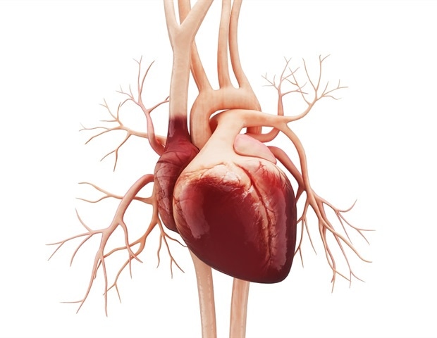
In a remarkable scientific achievement, researchers at UCL and the Francis Crick Institute have captured the first-ever real-time images of heart formation within a living mouse embryo. This innovative approach has unveiled the origins of cardiac cells, providing critical insights into the heart’s development. Understanding this process is vital for advancing cardiac research and potential treatments for heart diseases.

The use of 3D imaging technology has revolutionized how scientists study embryonic development. By enabling real-time observation, researchers can now witness the intricate processes that shape the heart. This breakthrough offers a window into the early stages of life, highlighting the complex interactions that govern organ formation.
Implications for Future Research
These findings pave the way for further exploration into cardiac health and disease. As scientists continue to unravel the mysteries of heart development, they can better understand congenital heart defects and other cardiovascular conditions. This research stands as a testament to the power of innovative imaging techniques in advancing our knowledge of biological processes.


