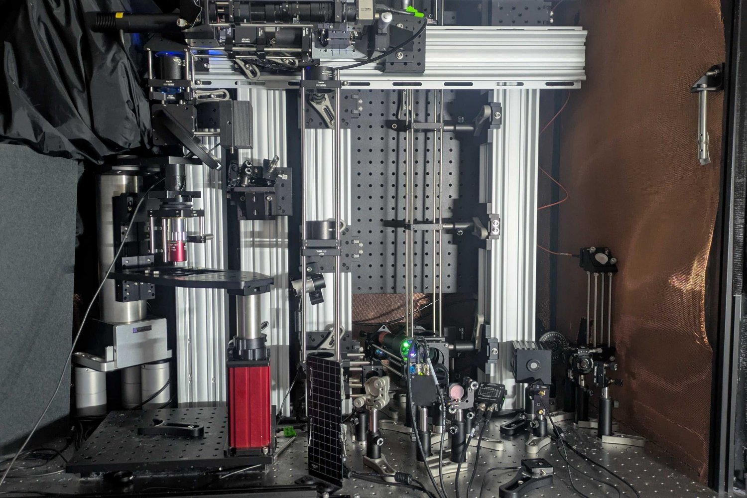
MIT researchers at the Picower Institute have developed a revolutionary imaging technology that enables scientists to peer deeper into living brain tissue than ever before. This cutting-edge microscope system uses sound waves to detect the molecular activity of individual cells, offering a new window into the complexities of the brain. By leveraging the power of sound, the system surpasses previous imaging limitations, providing single-cell resolution even at unprecedented depths within brain tissue.
Revolutionizing Brain Research
With this new technology, neuroscientists can observe the intricate workings of living brain cells in their natural environment. This advancement paves the way for deeper understanding of brain diseases, neural activity, and how individual cells interact. The ability to see such detail in live brain tissue could accelerate breakthroughs in treating conditions like Alzheimer’s, epilepsy, and other neurological disorders.
What Sets This Imaging System Apart?
Unlike traditional microscopes, which often struggle with light penetration and resolution at greater depths, the MIT team’s system combines advanced optics with acoustic techniques. This fusion allows for clearer, more detailed images of living cells, even far beneath the brain’s surface.
Sources:
Source